Cluster of DifferentiationCluster of Differentiation refers to a system of organizing and naming a molecule of antigen present on the outer surface of a cell membrane. [15] The abbreviation CD followed by a series of numbers is used to denote the presence of a structure to which specific antibodies bind. For example, CD4 refers to specific molecules found on T lymphocytes. However, these diminish in the presence of HIV, as this virus attacks the immune system. [15] 
Above is a picture of the CD4 molecule Click here to view entry in Protein Data Bank This nomenclature was developed in 1982 in the First International Workshop on Human Leukocyte Differentiation Antigens. The goal of this group of international immunologists was to define receptors and complex proteins to which antibodies could bind. In particular, they were focusing on cancer and its related antibodies. [15] |
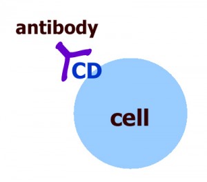 Above is a diagram demonstrating how an antibody binds to an antigen, or a Cluster of differentiation 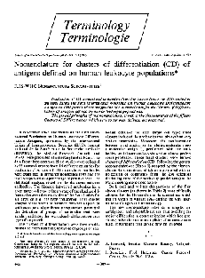 Above is a document from the First International Workshop on Human Leukocyte Differentiation Workshop on Human Leukocyte Antigens. Click here to view the document in a new window. |
Immunophenotyping
Immunophenotyping refers to the procedure by which cells are identified according to the molecules on their surfaces, especially clusters of differentiation. It is used to identify the proteins expressed by cells, and can be used to diagnose cancer and other diseases. [7]
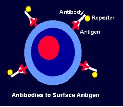 Above is a diagram demonstrating how antibodies, which are attached to reporters, bind to antigens. Below is the Immunofluorescence image of Giardia lamblia cysts 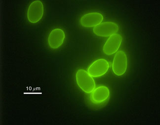 Below is a diagram explaining Flow Cytometry 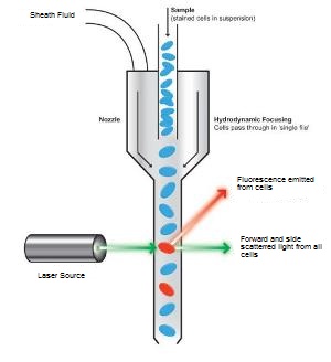
| How it works:A serum of antibodies is poured over a tissue sample. If the corresponding antigens are present on the cells, they bind with the antibodies, which also are attached to "reporters" which help to identify the antibodies. Next, the tissue is washed to remove any antibodies that may have bonded non-specifically. Finally, there are several ways to indicate the presence of the antibodies and antigens. They include immunofluorescence, flow cytometry, and immunohistochemistry.[7] Immunofluorescence: When a specific frequency of light is shone on the tissue, the reporter can be made to fluoresce. The presence of fluorescent color would then indicate that the antibody, and thus the antigen, is present in the tissue, meaning that the cell would be positive for the cluster of differentiation being identified.[7] Flow Cytometry When the tissue is suspended in liquid, a machine named a flow cytometer can be used to identify the reporters on the antibiotics. A thin stream of the solution is passed through a laser so that the cells can be exposed to the laser individually. According to the diffraction and if the reporters fluoresce, the cluster of differentiation can be identified. [7] Immunohistochemistry Sometimes, the antibody is attached to an enzyme. Rather than exposing it to light to cause it to fluoresce, the corresponding substrate is introduced. The enzyme then causes the substrate to change color and precipitate.[7] See the video to the right for a recap and explanation of flow cytometry. This video is a documentary done by a student working at the University of Utah. |